✨ This article was AI edited. Editorial responsibility: Interconnect-Diabetes.eu.
Produced with AI. This article was artificially generated. Editorial responsibility: Interconnect-Diabetes.eu.
Diabetic foot ulcers (DFUs) are among the most serious, debilitating, and costly complications of diabetes mellitus. Globally, approximately 15% to 25% of individuals diagnosed with diabetes will develop a foot ulcer during their lifetime. What begins as a minor blister or undetected abrasion from tight footwear can rapidly progress to deep-tissue infection, osteomyelitis, and lower-extremity amputation without timely intervention. Understanding the pathogenesis, Wagner classification stages, offloading modalities, advanced wound dressing selection, and multidisciplinary healing protocols empowers patients and clinicians to prevent tissue loss and achieve rapid ulcer resolution.
Clinical Highlights & Practice Essentials
- The Triad of Ulceration: Neuropathy (sensory loss), vascular disease (ischemia), and biomechanical pressure deformity constitute the primary triad leading to skin breakdown.
- Wagner Staging System: Ulcers are graded from Stage 0 (intact pre-ulcerative skin) to Stage 5 (extensive gangrene involving the entire foot), guiding surgical and antimicrobial therapy.
- Total Contact Casting (TCC): Non-removable total contact casting remains the gold-standard mechanical offloading modality, reducing peak plantar pressure by over 80%.
- Advanced Wound Care Therapies: Hyperbaric oxygen therapy (HBOT), cellular and tissue-based products (CTPs), and negative pressure wound therapy (NPWT) accelerate granulation in non-healing wounds.
Pathophysiology: How Diabetic Foot Ulcers Develop
The progression of a diabetic foot ulcer involves a complex interplay of systemic and local factors:
- Sensory Neuropathy: Loss of protective sensation (LOPS) prevents the patient from feeling pain, heat, or mechanical friction. Micro-trauma goes unnoticed and unaddressed.
- Motor & Autonomic Neuropathy: Motor nerve damage causes intrinsic foot muscle atrophy, resulting in structural claw toes and prominent metatarsal heads. Autonomic neuropathy leads to anhidrosis (absence of sweating), causing hyperkeratotic fissures and cracking.
- Peripheral Arterial Disease (PAD): Microvascular dysfunction and macrovascular arterial stenosis reduce capillary perfusion, depriving healing tissues of oxygen, nutrients, and circulating immune cells.
- Impaired Inflammatory Cascade: Chronic hyperglycemia induces excessive matrix metalloproteinase (MMP) activity, degrades extracellular matrix proteins, and impairs neutrophil phagocytosis, stalling wounds in a persistent inflammatory phase.
Wagner Ulcer Classification & Staging Matrix
| Wagner Grade | Clinical Presentation | Tissue Depth Involved | Recommended Clinical Intervention |
|---|---|---|---|
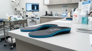
| Grade 0 | Pre-ulcerative lesion, healed ulcer, or bony deformity | Intact epidermis | Preventive custom orthotics, callus debridement, patient education |
| Grade 1 | Superficial ulcer without subcutaneous involvement | Epidermis and dermis only | Sharp debridement, moisture-retentive dressing, pressure offloading |
| Grade 2 | Deep ulcer penetrating to tendon, capsule, or bone | Subcutaneous tissue, tendon sheath | Surgical debridement, targeted oral/IV antibiotics, total contact casting |
| Grade 3 | Deep ulcer with abscess, osteomyelitis, or joint sepsis | Bone cortex and marrow space | Urgent surgical resection, bone biopsy, long-term targeted IV antibiotic therapy |
| Grade 4 | Localized gangrene involving toes or forefoot | Necrotic soft tissue and bone | Urgent vascular revascularization (angioplasty/bypass) and partial amputation |
| Grade 5 | Extensive gangrene involving entire foot | Full thickness necrosis of foot | Emergency major limb amputation (below-knee or above-knee) to prevent systemic sepsis |
Gold-Standard Clinical Healing Protocols
1. Sharp Surgical Debridement (TIME Principle)
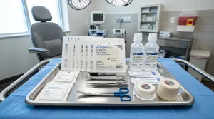
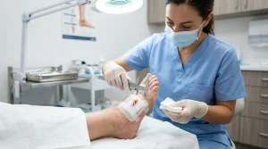
Regular conservative sharp debridement (CSWD) using a sterile scalpel or curette removes necrotic non-viable tissue, reduces bacterial bioburden, and disrupts chronic wound biofilms. By converting a stagnant chronic wound bed into an acute healing environment, debridement stimulates platelet-derived growth factors (PDGF) and fibroblast proliferation.
2. Mechanical Pressure Offloading
Without adequate mechanical offloading, no topical therapy or dressing will successfully heal a neuropathic plantar ulcer. The International Working Group on the Diabetic Foot (IWGDF) recommends non-removable knee-high devices (Total Contact Casts or locked walker boots) as the first-line standard of care. Removable cast boots and surgical shoes offer lower compliance and significantly slower healing rates.
3. Advanced Wound Dressings Selection
- Hydrogels: For dry, necrotic wound beds requiring autolytic debridement and moisture donation.
- Alginates & Hydrofibers: High-absorption calcium alginate dressings for heavily exuding wounds that form a cohesive gel upon contact with fluid.
- Silver Antimicrobial Dressings: Ionic silver or silver sulfadiazine foams for wounds exhibiting high bacterial bioburden or localized signs of critical colonization.
- Collagen & Extracellular Matrix (ECM) Scaffolds: Matrix-binding dressings that inhibit destructive matrix metalloproteinases and guide cellular re-epithelialization.
Prevention Protocols: Daily Foot Inspection & Podiatric Routine
- Mirror-Assisted Daily Inspection: Inspect all plantar surfaces, heels, and interdigital spaces under direct bright light for blisters, erythema, or maceration.
- Proper Moisture Management: Apply urea-based moisturizers daily to the heels and dorsum of the foot, but never between the toes to avoid interdigital fungal infection.
- Never Walk Barefoot: Always wear supportive, seamless diabetic shoes or structured indoor slippers with orthopedic footbeds.
- Monofilament Sensory Screening: Undergo routine annual 10g Semmes-Weinstein monofilament sensory testing and Ankle-Brachial Index (ABI) vascular assessment with a podiatrist.
Frequently Asked Questions (FAQ)
How long does it take for a diabetic foot ulcer to heal?
With optimal mechanical offloading, glycemic management (HbA1c <7.0%), infection control, and professional wound care, uncomplicated superficial ulcers typically heal within 6 to 12 weeks. Deep or ischemic wounds may require 4 to 6 months and specialized adjunctive therapies.
Can soaking feet in Epsom salt help heal a diabetic ulcer?
No. Podiatrists strictly forbid soaking feet in hot water or Epsom salts. Soaking causes skin maceration, breaks down fragile epidermal barriers, and risks severe thermal burns due to reduced sensory nerve perception.
When should a patient seek emergency medical attention for a foot wound?
Immediate emergency medical evaluation is required if you observe spreading redness around the wound, red streaks up the leg, fever or chills, foul-smelling purulent drainage, blackened necrotic tissue, or sudden severe glycemic spikes.
Get our latest guides, news, and insights highlighted in your Google Search & AI Overviews.