✨ This article was AI edited. Editorial responsibility: Interconnect-Diabetes.eu.
Produced with AI. This article was artificially generated. Editorial responsibility: Interconnect-Diabetes.eu.
Diabetic wound care is the clinical practice of assessing, debriding, offloading, and dressing cutaneous injuries in diabetic patients. Impaired by neuropathy and peripheral arterial disease, diabetic wounds heal through four phases (hemostasis, inflammation, proliferation, remodeling) and require sterile saline irrigation, moist wound healing, and offloading to prevent amputation.
Every twenty seconds, somewhere in the world, a lower limb is lost as a consequence of diabetes mellitus. More than 80% of these tragic amputations begin with a simple, preventable diabetic wound—a harmless blister, a cracked heel, or an unnoticed abrasion beneath a callused metatarsal head. In individuals living with diabetes, the combination of loss of protective sensation (LOPS), microvascular sclerosis, and diminished phagocytic immune response transforms minor tissue damage into chronic, non-healing ulcers that threaten life and limb.
Understanding the essential clinical stages of wound healing, recognizing early infection warning signs, mastering proper home first aid protocols, and utilizing advanced bioactive dressings are indispensable skills for diabetic patients, family caregivers, and healthcare providers.
The Physiology of Delayed Diabetic Wound Healing
Normal dermal wound healing proceeds through a synchronized, overlapping four-stage physiological cascade: hemostasis, inflammation, proliferation, and tissue remodeling. In patients suffering from chronic hyperglycemia, this biological progression becomes completely derailed:
| Healing Phase | Normal Wound Biology | Diabetic Pathophysiological Impairment | Clinical Manifestation |
|---|---|---|---|
| 1. Hemostasis | Immediate platelet plug formation; fibrin clot deposition; cytokine release (PDGF, TGF-beta) | Hyper-aggregable platelets; altered fibrin clot structure; endothelial dysfunction | Impaired microvascular clot dynamics; compromised initial signaling cascade |
| 2. Inflammation | Neutrophil and macrophage infiltration clearing bacteria and cellular debris (Days 1–4) | Impaired neutrophil chemotaxis; persistent M1 pro-inflammatory macrophage phenotype; elevated MMP-8 and MMP-9 | Chronic arrest in inflammatory phase; destruction of newly synthesized extracellular matrix |
| 3. Proliferation | Granulation tissue formation; angiogenesis; fibroblast migration; re-epithelialization | Blunted VEGF response; cellular senescence of fibroblasts; defective collagen deposition | Pale, non-granulating wound bed; failure of epithelial edge migration (“epibole”) |
| 4. Remodeling | Type III collagen replaced by organized Type I collagen; wound contraction (Months) | Accumulation of Advanced Glycation End-products (AGEs); cross-linking defects | Weak scar tensile strength (rarely exceeding 60% of original dermal strength); frequent recurrence |
Early Warning Signs: Recognizing Local & Deep-Tissue Infection
Due to peripheral neuropathy, pain is notoriously absent in up to 70% of infected diabetic wounds. Clinicians and patients must rely on objective physical markers to detect infection before it invades deep tendon sheaths and periosteum:
Superficial Infection Indicators (The Secondary Signs)
- Erythema (Periwound Redness): An erythematous halo extending less than 2 cm from the ulcer margin.
- Local Hyperthermia: An infrared dermal skin temperature increase of >2.2°C (4°F) compared to the contralateral foot.
- Exudate Alteration: Transition from clear serous fluid to thick purulent drainage with a pungent, foul odor.
- Friable Granulation Tissue: Wound bed that bleeds excessively with gentle probing or contact.
Deep-Tissue & Systemic Emergency Warnings
Immediate hospitalization and surgical consultation are required if any of the following symptoms emerge:
- Cellulitis >2 cm: Rapidly expanding redness ascending the ankle or lower leg.
- Positive Probe-to-Bone Test: Palpating hard, gritty cortical bone with a sterile metallic blunt probe through the wound bed (positive predictive value >85% for osteomyelitis).
- Subcutaneous Crepitus: Crackling sensation beneath the skin indicative of gas gangrene produced by anaerobic organisms (e.g., Clostridium perfringens).
- Systemic Inflammatory Response: Fever (>38°C), persistent tachycardia, leukocytosis, or sudden unexplained hyperglycemia resistant to insulin correction.
Step-by-Step Clinical Home First Aid Protocol for Diabetic Wounds
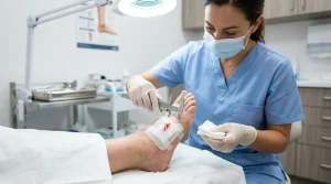
When an acute abrasion, tear, or puncture is discovered on a diabetic foot, follow this strict sterile first aid protocol:
Step 1: Cleansing and Irrigation
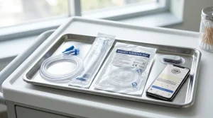
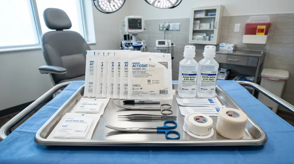
Gently irrigate the wound with sterile 0.9% normal saline or lukewarm potable tap water using a low-pressure bulb syringe. Avoid cytotoxic chemical antiseptics such as hydrogen peroxide, isopropyl alcohol, or undiluted povidone-iodine, which destroy fragile migrating keratinocytes and delay healing.
Step 2: Hemostasis and Inspection
Apply gentle pressure with sterile woven gauze. Once bleeding ceases, inspect the depth under bright illumination with a magnifying loupe, taking note of foreign bodies or tracking sinus tracts.
Step 3: Moisture-Retentive Dressing Selection
Apply an advanced bioactive dressing tailored to exudate levels. For dry or low-exudate wounds, apply a sterile amorphous hydrogel covered with a semi-permeable polyurethane foam dressing. For moderately to heavily exuding wounds, utilize a calcium alginate or hydrofiber pad that absorbs drainage without sticking to the wound bed.
Step 4: Immediate Mechanical Offloading
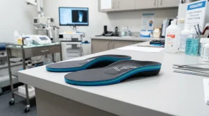
Never walk barefoot or apply standard tight footwear over a fresh wound. Transfer immediately into a cushioned surgical post-op shoe or non-weight-bearing status, and contact your specialist podiatry foot clinic within 24 hours.
Nutritional Optimization for Accelerated Wound Healing
Dermal tissue regeneration demands a substantial metabolic investment. When an open wound is present, systemic catabolism increases dramatically, requiring targeted macro- and micronutrient support. Dietary protein intake should be increased to 1.25 to 1.5 grams per kilogram of body weight per day to provide essential amino acids (particularly L-arginine and L-glutamine) necessary for fibroblast proliferation and collagen cross-linking.
Specific micronutrients also play decisive enzymatic roles: Vitamin C acts as an obligate cofactor for prolyl and lysyl hydroxylase in collagen triple-helix stabilization, while elemental Zinc (15 to 30 mg daily) serves as a catalyst for DNA polymerase and matrix metalloproteinase regulation. Supplementing with therapeutic doses of bioactive antioxidants combats localized oxidative stress, enabling more rapid granulation tissue formation in refractory diabetic foot ulcers.
The Role of Podiatric Offloading Modalities in Daily Life
Mechanical offloading remains the single most decisive factor predicting diabetic ulcer resolution. Without eliminating vertical ground reaction forces and shear friction, advanced wound dressings and growth factors cannot stimulate re-epithelialization. Total Contact Casting (TCC) remains the undisputed gold standard, reducing peak plantar pressure by redistributing body mass across the entire lower leg. For patients unable to tolerate rigid casts, removable cast walkers (RCWs) rendered non-removable with a security strap or specialized felted foam padding provide effective alternative pressure redistribution.
Frequently Asked Questions
Should a diabetic wound be left open to “air out”?
No. Modern wound biology conclusively proves that open exposure leads to dermal dehydration, cellular death, and bacterial contamination. Moist wound healing accelerates epithelialization by up to 40% compared to dry scab formation.
Can I soak my diabetic foot wound in an Epsom salt foot bath?
No. Foot baths cause cutaneous maceration (softening and waterlogging of healthy skin), which breaks down tissue barriers and allows bacterial pathogens to penetrate deeper into subcutaneous compartments.
What is hyperbaric oxygen therapy (HBOT) for diabetic wounds?
HBOT involves breathing 100% medical oxygen in a pressurized hyperbaric chamber (2.0–2.4 atmospheres). This super-saturates blood plasma with dissolved oxygen, stimulating neo-vascularization and enhancing bacterial destruction in severely hypoxic, ischemic diabetic ulcers.
Get our latest guides, news, and insights highlighted in your Google Search & AI Overviews.