✨ This article was AI edited. Editorial responsibility: Interconnect-Diabetes.eu.
Produced with AI. This article was artificially generated. Editorial responsibility: Interconnect-Diabetes.eu.
Types of diabetic wounds include neuropathic ulcers, ischemic ulcers, neuro-ischemic lesions, and infectious wounds such as diabetic cellulitis or osteomyelitis. Commonly classified using the Wagner Ulcer Grade (0–5) or University of Texas staging system, they stem from peripheral neuropathy, peripheral arterial disease (PAD), and microvascular impairment requiring specialized offloading and debridement.
Chronic wounds represent one of the most severe, debilitating complications associated with long-term diabetes mellitus. Across the globe, an estimated 19% to 34% of individuals diagnosed with diabetes will develop a diabetic foot ulcer (DFU) during their lifetime. Due to the triad of peripheral sensory neuropathy, peripheral vascular insufficiency, and compromised immunocompetence, minor abrasions or calluses can rapidly degrade into deep tissue necrosis, limb-threatening infections, and surgical amputations.
Understanding the distinct physiological classifications, diagnostic criteria, clinical stages, and evidence-based therapeutic interventions for diabetic wounds is paramount for effective patient care and limb salvage.
Etiological Classifications of Diabetic Wounds
Diabetic wounds do not present uniformly; their clinical presentation, pain profile, bleeding potential, and healing trajectory depend fundamentally upon the underlying vascular and neurological pathophysiology.
| Wound Type | Primary Cause | Typical Location | Appearance & Margins | Pain Perception | Vascular Status |
|---|---|---|---|---|---|
| Neuropathic Ulcer | Sensory/autonomic nerve fiber death; sustained pressure | Plantar surface of metatarsal heads, heel, plantar hallux | “Punched-out” round borders; surrounded by thick hyperkeratotic callus; pink granulation bed | Painless or diminished sensation (loss of protective threshold) | Palpable peripheral pulses; warm foot; normal capillary refill |
| Ischemic Ulcer | Macrovascular Atherosclerosis (Peripheral Artery Disease) | Tips of toes, lateral foot borders, over bony prominences, interdigital spaces | Irregular, poorly defined margins; pale, necrotic, or black eschar; minimal granulation | Intense pain, often worsening with elevation or at night (rest pain) | Diminished or absent dorsalis pedis / posterior tibial pulses; cool, pale, shiny skin; loss of hair |
| Neuro-Ischemic Ulcer | Combined sensory neuropathy and arterial insufficiency | Margin of the foot, heels, and dorsal toe joints | Mixed borders, variable necrotic slough, thin hyperkeratosis | Variable: blunted due to neuropathy, yet intermittently painful | Compromised pulses; ABI < 0.8; sluggish capillary refill |
| Infectious Diabetic Lesion | Secondary bacterial invasion (Staph, Strep, Pseudomonas, anaerobes) | Extending from pre-existing ulcer, fissures, or puncture sites | Erythematous, edematous halo (>2 cm indicates severe infection), purulent exudate, foul odor | Sudden local pain or constitutional malaise, fever | Septic thrombosis can induce acute ischemic necrosis |
Clinical Staging Systems: Wagner vs. University of Texas
Standardized grading systems allow multidisciplinary wound care teams to determine clinical urgency, establish amputation risk, and track wound healing progression.
The Wagner Ulcer Classification System
Historically the most widespread metric, the Wagner scale grades wounds based primarily on anatomical depth and the presence of gangrene:
- Grade 0: Intact skin with high-risk structural deformities (e.g., Charcot arthropathy, prominent metatarsal heads, severe claw toes).
- Grade 1: Superficial ulceration involving the full thickness of the epidermis and dermis, but not penetrating into tendon, capsule, or bone.
- Grade 2: Deep ulcer penetrating to tendon, ligament, joint capsule, or deep fascia, without active abscess or osteomyelitis.
- Grade 3: Deep ulcer complicated by osteitis, abscess formation, tenosynovitis, or osteomyelitis.
- Grade 4: Localized gangrene involving the forefoot, toes, or heel.
- Grade 5: Extensive gangrene involving the entire foot requiring major surgical amputation.
The University of Texas (UT) Staging System
While the Wagner scale assesses depth and gangrene, the University of Texas Wound Classification matrix is widely considered clinically superior because it cross-references depth (Grades 0–3) against ischemic and infectious status (Stages A–D). For instance, a Stage B Grade 2 wound represents a deep tendon-level wound with active infection, while Stage D Grade 2 indicates deep tissue involvement with combined infection and severe ischemia—carrying a dramatically higher risk of non-healing.
Core Pillars of Diabetic Wound Management & Limb Salvage
Achieving successful wound closure in diabetic patients requires a synchronized multidisciplinary strategy based on four cardinal principles:
1. Mechanical Offloading
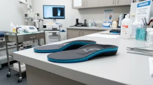
Continuous mechanical friction and perpendicular sheer pressure prevent cellular migration and destroy newly formed capillary buds. The gold standard for offloading plantar neuropathic ulcers is the Total Contact Cast (TCC) or non-removable pneumatic walking boot. By redistributing load across the entire calf and plantar arch, offloading reduces peak plantar pressures at the ulcer site by up to 85%.
2. Surgical & Sharp Debridement
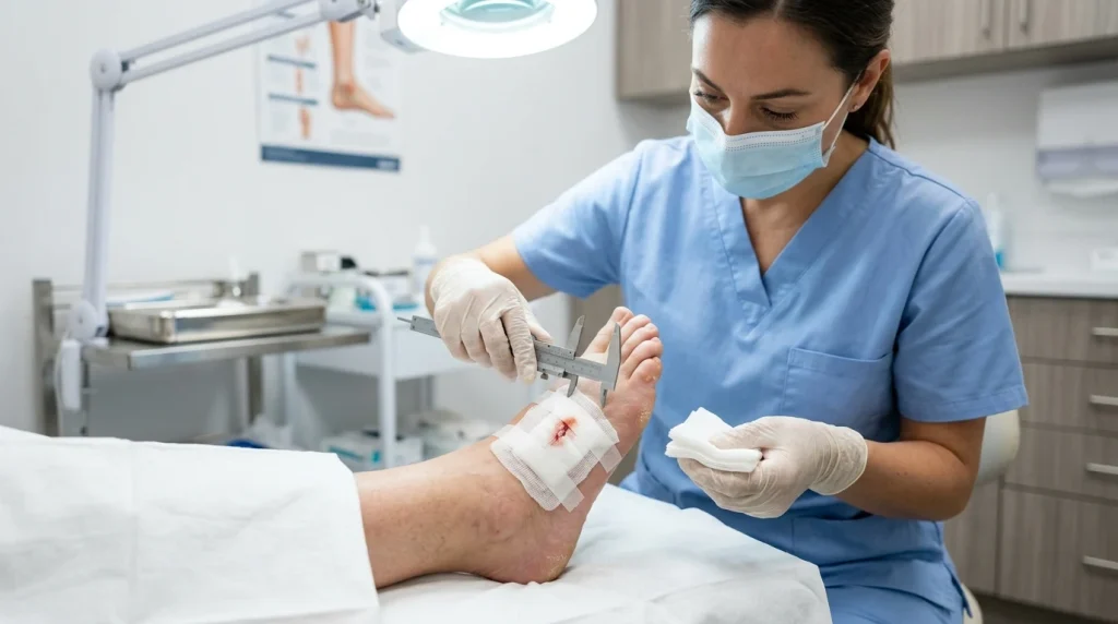
Chronic diabetic wounds contain senescent cells, bacterial biofilms, and necrotic debris that produce inflammatory cytokines (MMP-8, MMP-9) inhibiting collagen synthesis. Regular sharp debridement converts a chronic, non-responsive wound bed back into an acute healing state, exposing healthy bleeding margins that stimulate endogenous growth factors.
3. Advanced Bioactive Dressings & Moisture Balance
Modern wound protocols avoid desiccating antiseptics like iodine in favor of advanced dressings:
- Alginates and Hydrofibers: Highly absorbent dressings engineered for heavily exuding wounds that turn into a soothing gel upon contact with wound fluid.
- Cadexomer Iodine or Silver Nanoparticle Foams: Deliver sustained antimicrobial action against resistant biofilms (such as MRSA) without cytotoxic damage to granulating fibroblasts.
- Collagen & Extracellular Matrix Matrices: Provide sacrificial substrates that absorb destructive matrix metalloproteinases, allowing the patient’s natural tissue scaffold to rebuild.
4. Vascular Revascularization
No wound can heal without oxygenated blood delivering immune cells, antibiotics, and nutrients. When an Ankle-Brachial Index (ABI) reads below 0.5, or transcutaneous oxygen pressure (TcPO2) falls below 30 mmHg, immediate referral to a vascular surgeon for endovascular angioplasty or surgical bypass grafting is vital before attempting aggressive local interventions.
When to Seek Emergency Medical Attention
Diabetic patients and caregivers should report immediately to an emergency medical department or specialist foot clinic if any of the following “red flag” symptoms appear:
- Rapidly spreading redness (cellulitis) extending more than 2 centimeters beyond the wound margin.
- New black, purple, or mottled skin discoloration indicative of tissue death (dry or wet gangrene).
- Crepitus (a crackling sensation under the skin upon palpation), which suggests gas-producing necrotizing fasciitis.
- Systemic signs of sepsis: unexplained fever, chills, confusion, or uncontrollable blood sugar spikes unresponsive to standard insulin administration.
Frequently Asked Questions
Why do diabetic wounds heal so slowly?
Hyperglycemia impairs white blood cell function (chemotaxis and phagocytosis), thickens capillary basement membranes reducing tissue perfusion, and increases oxidative stress, locking the wound in a persistent inflammatory phase.
Can an infected diabetic ulcer be treated with oral antibiotics alone?
Mild superficial infections may respond to targeted oral antibiotics, but moderate-to-severe infections involving deep structures, tendon, or bone require surgical debridement, tissue culturing, offloading, and often intravenous antibiotic therapy.
What is Charcot foot and how does it relate to diabetic wounds?
Charcot foot is a severe neuroarthropathy characterized by micro-fractures, bone reabsorption, and joint collapse (often resulting in a “rocker-bottom” sole) caused by severe neuropathy. The abnormal bony prominence dramatically increases localized pressure, frequently leading to deep, refractory ulcerations.
Get our latest guides, news, and insights highlighted in your Google Search & AI Overviews.