✨ This article was AI edited. Editorial responsibility: Interconnect-Diabetes.eu.
Produced with AI. This article was artificially generated. Editorial responsibility: Interconnect-Diabetes.eu.
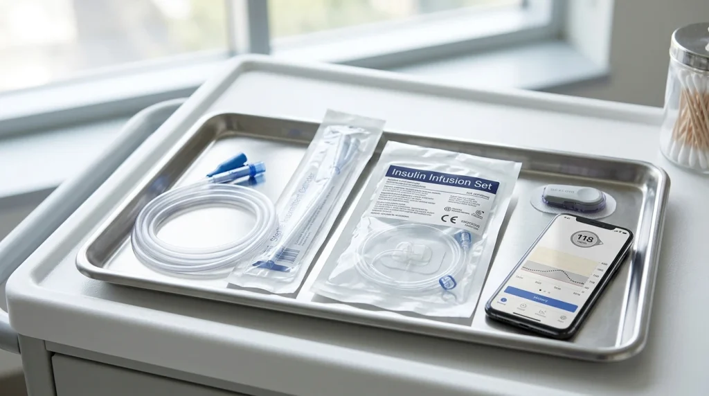
A diabetes catheter typically refers to either a subcutaneous insulin infusion cannula connecting an external insulin pump to fatty tissue, or a urinary catheter managed in diabetic patients. Both medical devices require meticulous hygiene protocols due to elevated risks of catheter-associated urinary tract infections (CAUTI), cannula occlusion, and subcutaneous lipohypertrophy.
In medical discourse, the term catheter carries multifaceted meanings for individuals managing diabetes. Most commonly, it denotes the soft cannula or steel needle component of continuous subcutaneous insulin infusion (CSII) pump systems. Concurrently, in clinical and acute hospital settings, it refers to indwelling (Foley) or intermittent urethral catheters used to manage diabetic neurogenic bladder—a common manifestation of diabetic autonomic neuropathy.
Whether navigating the daily mechanics of automated insulin delivery (AID) infusion sets or managing urinary tract catheterization under hyperglycemic conditions, proper device handling, rotation protocols, and infection surveillance are foundational to preventing acute metabolic crises and systemic complications.
Insulin Pump Infusion Sets: Anatomy, Types & Rotation Mechanics
For pump users, the infusion catheter is the physiological bridge between the automated insulin reservoir and subcutaneous adipose tissue. Selecting the appropriate catheter configuration directly influences insulin absorption rates, glycemic variability, and cutaneous tolerance.
| Catheter Type | Material | Insertion Angle | Cannula Length | Ideal Patient Profile | Key Clinical Consideration |
|---|---|---|---|---|---|
| Standard 90-Degree Soft Cannula | Flexible Teflon / Polyurethane | 90° (Straight perpendicular) | 6 mm, 9 mm | Average subcutaneous fat depth; children; general pump users | Comfortable during physical movement; susceptible to kinking or bending under muscular tension |
| Angled Soft Cannula | Flexible Teflon | 20° to 45° (Oblique insertion) | 13 mm, 17 mm | Lean, athletic individuals; pregnant women; patients with minimal abdominal fat | Allows precise placement into shallow fat layers; requires manual or angled inserter |
| Stainless Steel Needle Cannula | Medical-grade Stainless Steel | 90° straight | 5.5 mm, 8.5 mm | Patients with Teflon allergies; history of unexplained kinks; scar tissue | Zero risk of cannula kinking; must be changed every 48 hours to minimize local inflammation |
| Extended-Wear Infusion Sets | Advanced polymer tubing and biocompatible adhesive | 90° perpendicular | 6 mm, 9 mm | Patients seeking reduced change frequency; active lifestyles | Engineered for up to 7 consecutive days of wear without insulin degradation or site reactions |
Troubleshooting Pump Catheter Occlusions & Lipohypertrophy
Unexplained, persistent hyperglycemia unresponsive to corrective insulin boluses is the hallmark sign of catheter failure. Subcutaneous cannulas can fail through several distinct mechanisms:
- Cannula Kinking: Often occurs during insertion when the introducer needle retracts prematurely, or when the cannula encounters dense subcutaneous fascial planes.
- Silent Insulin Crystallization: Body heat combined with turbulent fluid flow inside microscopic tubing can cause insulin hexamers to dissociate into fibrils, creating micro-clogs that trigger pump “Occlusion Detected” alarms.
- Lipohypertrophy: Repeatedly inserting catheters into the same anatomical quadrant causes adipocytes to hypertrophy and synthesize dense fibrous tissue. Delivering insulin into a lipohypertrophic nodule causes erratically delayed absorption (up to 3–4 hours), resulting in severe postprandial spikes followed by unpredictable late hypoglycemia.
- Rotation Protocol: Clinicians mandate a minimum separation of 2 inches (5 cm) between consecutive catheter insertion sites, systematically cycling across the abdomen, upper buttocks, flanks, and anterior-lateral thighs.
Diabetic Neurogenic Bladder & Urinary Catheterization Care
Prolonged hyperglycemia can inflict irreversible microvascular damage upon the parasympathetic and sympathetic nerves innervating the detrusor muscle and internal urethral sphincter—a pathological state known as diabetic cystopathy or neurogenic bladder.
In its early stages, diabetic cystopathy impairs bladder sensation, causing patients to fail to perceive full bladder fullness. Over years, this progresses to chronic urinary retention, hydronephrosis, and overflow incontinence, necessitating either Clean Intermittent Catheterization (CIC) or the placement of an indwelling Foley catheter.
The Heightened Risk of Catheter-Associated Urinary Tract Infections (CAUTI)
Individuals with diabetes face an approximately twofold to fourfold higher risk of developing severe, bacteremic CAUTIs compared to non-diabetic cohorts. Several pathophysiological factors contribute to this susceptibility:
- Glucosuria as a Microbial Culture Medium: When renal thresholds (>10 mmol/L or ~180 mg/dL) are exceeded, excess glucose is excreted in the urine, providing an optimal growth medium for uropathogens like Escherichia coli, Klebsiella pneumoniae, and opportunistic fungi (Candida albicans).
- Impaired Neutrophil Phagocytosis: Hyperglycemia paralyzes polymorphonuclear leukocytes, blunting their ability to migrate toward and eliminate bacterial biofilms adhering to silicon and latex catheter lumens.
- Ascending Pyelonephritis Risk: Due to vesicoureteral reflux and incomplete voiding, localized bladder infections can rapidly migrate up the ureters to cause acute emphysematous pyelonephritis—a life-threatening urological emergency.
Evidence-Based Guidelines for Safe Catheter Maintenance
Whether caring for an insulin delivery cannula or a urinary catheter system, adherence to strict clinical maintenance guidelines protects against sepsis and tissue necrosis:
- Aseptic Site Preparation: Before inserting or manipulating any catheter apparatus, perform a 30-second hand scrub with antimicrobial soap. Disinfect the skin surface using 2% chlorhexidine gluconate in 70% isopropyl alcohol and allow it to dry completely for at least 2 minutes to prevent chemical irritation and bacterial inoculation.
- Tubing Security & Tension Relief: Secure tubing using tension-relief loops (such as StatLock devices for urinary catheters or dedicated medical adhesives for pump lines). This prevents repetitive micro-trauma from pulling or snagging on clothing.
- Closed Drainage Integrity: For indwelling urinary catheters, ensure the drainage bag remains strictly below the level of the bladder at all times to eliminate gravitational backflow of stagnant, colonized urine.
- Strict Exchange Intervals: Never exceed recommended manufacturer wear durations. Standard Teflon pump catheters should be replaced every 48 to 72 hours; steel needles every 48 hours; urinary catheters per urologist scheduling.
Summary: Vigilance Ensures Safety
Catheters are indispensable life-sustaining medical technologies in diabetes care. By respecting site rotation rules for continuous subcutaneous insulin infusion and adhering to rigorous aseptic protocols for urinary drainage systems, patients can harness the therapeutic power of catheter technology while minimizing infectious and mechanical hazards.
Frequently Asked Questions
How do I know if my insulin pump catheter is bent or failed?
Symptoms include unexpected high blood glucose readings that do not decrease 1–2 hours after a correction bolus, the scent of insulin around the infusion site, wetness or leakage under the adhesive tape, or recurring pump occlusion alarms.
Is intermittent catheterization better than an indwelling catheter for diabetics?
Yes. Clean Intermittent Catheterization (CIC) significantly reduces the incidence of chronic biofilm formation and CAUTIs compared to continuous indwelling Foley catheters, preserving bladder wall tone and reducing systemic infection rates.
Can having a catheter cause ketones to appear?
If an insulin catheter becomes occluded, detached, or bent, the body stops receiving basal insulin. In patients with type 1 diabetes, absolute insulin deficiency can trigger diabetic ketoacidosis (DKA) in as little as 3 to 4 hours, accompanied by rapid ketone accumulation.
Get our latest guides, news, and insights highlighted in your Google Search & AI Overviews.